
Free PyMOL and Molecular Visualization Certification Assessment | StemSkills Lab
Skip to content

Free PyMOL and Molecular Visualization Certification Assessment

Test your knowledge of PyMOL and molecular visualization: selections, structure alignment, publication-quality figures, and protein-ligand interaction diagrams. Pass at 70% to earn a verifiable StemSkills certificate. Download it as a PDF and add it to your LinkedIn profile. It is free.
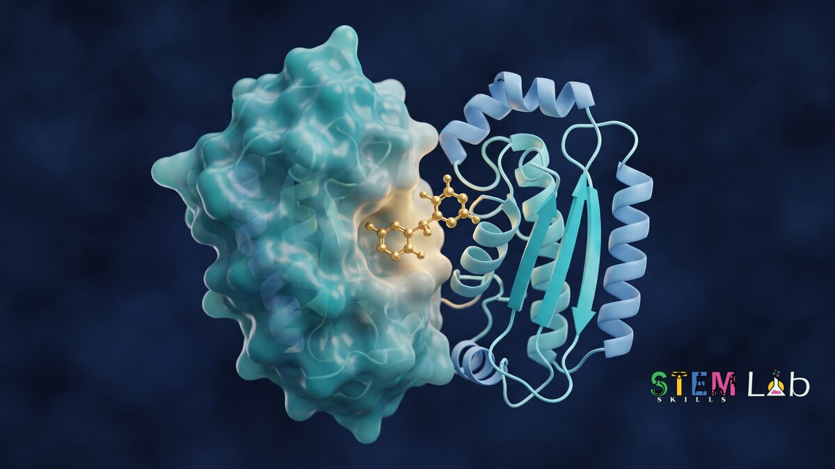
PyMOL and Molecular Visualization certification assessment

1
Take the quiz
20 questions on PyMOL and molecular visualization. A 20-minute timer runs once you start. No sign-up needed to start.
2
Sign in to see your result
One click with Google. Your score is saved to your account so you can see whether you passed.
3
Pass? Get certified
Score 70% or more and download your verifiable certificate, then add it to LinkedIn.
time left:
:
:

PyMOL and Molecular Visualization Certification Assessment

Submit quiz
Once you submit, you will no longer be able to change your answers. Are you sure you want to submit the quiz?

Free certification assessment. Pass at 70% to earn a verifiable StemSkills certificate.

1.
In PyMOL, the command fetch 1hsg:
Saves the current session as 1hsg.pse
Renders an image called 1hsg.png
Downloads PDB entry 1HSG and loads it into the session
Deletes the object named 1hsg
2.
Which command removes water molecules from the loaded structure?
hide waters
clean water
remove solvent
delete all
3.
In PyMOL selection syntax, resn and resi select by:
Chain and segment
Atom name and element
Residue name and residue number
Secondary structure and B-factor
4.
The ligand in 1HSG has the residue name MK1. Which command selects every complete residue with an atom within 4 Å of it?
select pocket, resn MK1
select pocket, resi 4
select pocket, byres (all within 4 of resn MK1)
select pocket, chain 4
5.
Two proteins share the same fold but very low sequence identity. The better PyMOL command to superimpose them is:
ray
align, because it ignores structure
fetch
super, because it does not depend on sequence alignment
6.
align reports an RMSD of 0.4 Å, but over far fewer atoms than the proteins contain. The reason is:
The proteins are identical
RMSD is always computed on one atom
PyMOL failed to load one protein
align runs outlier-rejection cycles, so the final RMSD covers only the atoms it kept
7.
The ray command in PyMOL:
Measures a distance
Ray-traces the scene to produce a high-quality image with shadows
Removes hydrogens
Rotates the camera by 90 degrees
8.
Which command writes a ray-traced 300 dpi image that is 2400 by 1800 pixels?
ray figure.png
save figure.png
show figure.png, 300
png figure.png, width=2400, height=1800, dpi=300, ray=1
9.
To export a ray-traced image with a transparent background, you set:
set ray_opaque_background, 0
set cartoon_transparency, 1
bg_color black
set transparency, 1
10.
Which command draws the polar contacts between a ligand selection lig and a protein selection prot?
color red, prot
distance hbonds, lig, prot, mode=2
zoom lig
show sticks, lig
11.
A figure labels a dashed line of 5.2 Å between a ligand oxygen and a protein nitrogen as a hydrogen bond. The best critique is:
Nitrogen cannot take part in hydrogen bonds
It is too long, because donor to acceptor distances in hydrogen bonds are typically about 2.5 to 3.5 Å
Distance is irrelevant to hydrogen bonds
Hydrogen bonds are usually longer than 6 Å, so it is too short
12.
To colour an AlphaFold model by its confidence in PyMOL you can run spectrum b, because:
PyMOL downloads confidence values from the internet
b selects beta sheets
b stands for backbone
AlphaFold files store pLDDT in the B-factor column
13.
A PyMOL file with the extension .pse is:
A script of commands
A coordinate file with atoms only
An image
A session file that keeps objects, representations, colours and the view
14.
You need two figures of different complexes from exactly the same camera angle. The reliable method is:
Use ray twice
Run get_view on the first scene and apply the printed matrix with set_view on the second
Rotate the second one by eye
Use zoom on both
15.
In a PDB format file, HETATM records hold:
Atoms of non-polymer groups such as ligands, ions and waters
The crystallographic resolution
Only hydrogen atoms
Only backbone atoms of the protein
16.
Very large structures such as ribosomes are distributed in PDBx/mmCIF rather than the legacy PDB format because the legacy format:
Only stores NMR data
Cannot store proteins
Has fixed-width columns that limit a file to 99,999 atoms and 62 chains
Is encrypted
17.
In UCSF ChimeraX, which command fetches and opens PDB entry 1HSG?
open 1hsg
get 1hsg
load 1hsg
fetch 1hsg
18.
Which tool produces a 2D schematic of a protein-ligand complex, with hydrogen bonds as dashed lines and hydrophobic contacts as spoked arcs?
BLAST
Open Babel
GROMACS
LigPlot+
19.
PLIP (Protein-Ligand Interaction Profiler) is used to:
Predict a structure from sequence
Convert file formats
Run molecular dynamics
Detect non-covalent protein-ligand interactions automatically from a structure file
20.
The correct way to credit PyMOL in a paper is to:
Cite "The PyMOL Molecular Graphics System", with the version, Schrödinger, LLC
Mention it only in the acknowledgements
Cite the Protein Data Bank instead
No credit is needed for open-source software
  • 1
  • 2
  • 3
  • 4
  • 5
  • 6
  • 7
  • 8
  • 9
  • 10
  • 11
  • 12
  • 13
  • 14
  • 15
  • 16
  • 17
  • 18
  • 19
  • 20
Ready to go beyond the assessment?
Our live, mentor-led cohort takes you hands-on through protein modelling, docking and molecular dynamics, finishing with a project you build yourself and can show on your CV and applications.

See the live cohort

See live workshops