
How to Predict Conformational B-Cell Epitopes

Conformational B-cell epitope prediction takes a 3D structure, not a sequence, and returns residues that sit together on the surface even when they are far apart in the chain. The three free servers are ElliPro, DiscoTope 3.0 and SEPPA 3.0. Each scores surface residues, clusters them in space, and ranks the resulting patches.
If you have already run a sequence-based predictor and are now holding a list of contiguous stretches, this is the step your earlier result could not perform. Our guide to predicting linear B-cell epitopes with BepiPred and ABCpred ends by telling you to move from linear prediction to a structure. This page is that move.
What you will find on this page
- Why a sliding window can never return a discontinuous epitope, proved on a real antibody complex
- What ElliPro, DiscoTope 3.0 and SEPPA 3.0 each measure, compared in one table
- The real parameters on each form, including the one DiscoTope setting students get wrong
- Whether these servers are valid on a designed multi-epitope construct, answered honestly
- A troubleshooting section built from real failures, including the residue-numbering trap
- FAQ
Why can a linear predictor never return a conformational epitope?
Because of how it reads the protein. A sequence-based predictor scans a window along the chain and scores the residues inside that window. Whatever it returns is therefore one contiguous run. An antibody does not bind a contiguous run. It presses onto a surface, and the residues under that footprint can come from anywhere in the chain.
The distinction is not new. Barlow, Edwards and Thornton set it out in 1986 in Nature, separating continuous from discontinuous antigenic determinants (Barlow DJ, Edwards MS, Thornton JM, Nature 1986;322:747-748). Most epitopes on native antigens are conformational, so a sequence-only result should always be described as a linear epitope prediction rather than as the epitope.
The proof, on a structure you can download
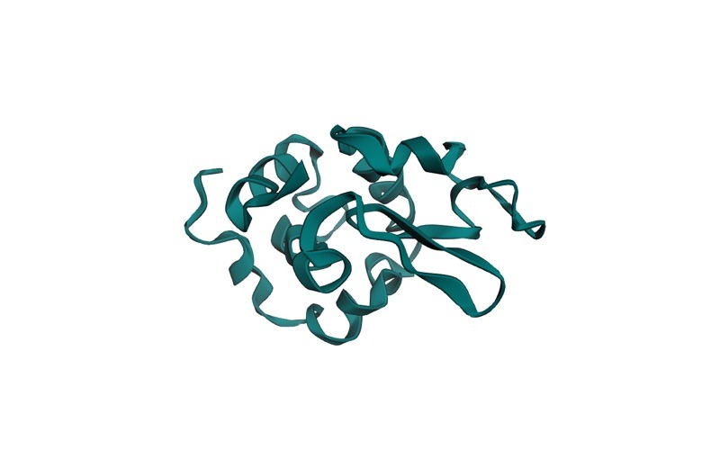
PDB entry 3HFM is titled “STRUCTURE OF AN ANTIBODY-ANTIGEN COMPLEX. CRYSTAL STRUCTURE OF THE HY/HEL-10 FAB-LYSOZYME COMPLEX”. It holds three chains, solved at 3.00 Angstrom resolution: H and L are the HyHEL-10 Fab, and chain Y is hen egg white lysozyme, 129 residues long. Because the complex is solved, the epitope is not a prediction here. It is a measurement.
Take every heavy atom of chain Y that sits within 4.0 Angstrom of any heavy atom of chain H or chain L, excluding hydrogens and keeping only the first alternate location. That gives exactly 17 antigen residues:
R14, H15, G16, N19, Y20, R21, W63, R73, L75, T89, N93, K96, K97, I98, S100, D101, G102
Those 17 residues form nine separate sequence segments: 14 to 16, 19 to 21, 63, 73, 75, 89, 93, 96 to 98, and 100 to 102. They are spread from residue 14 to residue 102 of a 129-residue protein. Loosen the cutoff to 5.0 Angstrom and you get 22 residues in eight segments, adding L17, D18, V99, N103 and A107.
Read that footprint again and the argument finishes itself. No window of 10 to 20 residues can contain nine segments spanning 88 residues of chain. A fixed-width predictor such as ABCpred is not performing badly on this antigen. It is being asked a question its input cannot express. You can reproduce the calculation on any complex: download the coordinate file, select the antigen chain, and keep the residues with at least one heavy atom inside your chosen distance of the antibody chains.
What do the three structure-based servers actually do?
All three start from coordinates and end with ranked surface patches, but they get there by different routes, and the route determines how much you should trust the number.
ElliPro is geometric. Its help page states that it “predicts linear and discontinuous antibody epitopes based on a protein antigen’s 3D structure” and that it “associates each predicted epitope with a score, defined as a PI (Protrusion Index) value averaged over epitope residues”. The protrusion index comes from fitting ellipsoids to the protein: the documentation explains that “the ellipsoid with PI = 0.9 would include within 90% of the protein residues with 10% of the protein residues being outside of the ellipsoid”, and that “Residues with larger scores are associated with greater solvent accessibility”. Discontinuous epitopes are then “clustered based on the distance R (in Angstrom between residue’s centers of mass)”. The method implements Thornton’s protrusion idea directly, which is why ElliPro’s own documented example includes myohemerythrin (2MHR).
The ElliPro paper is open access and reports a real benchmark: “In comparison with six other structure-based methods that can be used for epitope prediction, ElliPro performed the best and gave an AUC value of 0.732, when the most significant prediction was considered for each protein”, and “the rank of the best prediction was at most in the top three for more than 70% of proteins and never exceeded five” (Ponomarenko et al., BMC Bioinformatics 2008;9:514). The same paper notes that ElliPro “is based on the geometrical properties of protein structure and does not require training”. That single clause explains both its strengths: it cannot overfit a training set, and it cannot learn from one either, which is why an AUC near 0.73 is a fair result rather than a disappointing one.
One thing to know before you cite the classic server: every ElliPro page now carries the notice “This tool has been integrated into the B Cell Prediction – Structure Input tool on our Next-Generation Tools site.” The classic server at tools.iedb.org/ellipro is still live and still has the readable documentation, while IEDB is moving the method to nextgen-tools.iedb.org.
DiscoTope 3.0 is learned, not geometric. It uses inverse folding latent representations, and its paper reports that it “maintains high predictive performance across solved, relaxed and predicted structures, alleviating the need for experimental structures and extending the general applicability of accurate B-cell epitope prediction by 3 orders of magnitude” (Hoie et al., Frontiers in Immunology 2024;15:1322712). For a student whose antigen has no crystal structure, that sentence is the whole reason to prefer it.
SEPPA 3.0 is the glycoprotein specialist. Its server describes itself as “spatial epitope prediction for protein antigens, particularly for N-linked glycoproteins” and as “the first algorithm considering the N-glycosylation sites”. Its paper reports that “The AUC value of 0.794 was obtained through 10-fold cross-validation on internal validation” and that “Independent testing on general protein antigens resulted in AUC of 0.740 with BA (balanced accuracy) of 0.657” (Zhou et al., Nucleic Acids Research 2019;47(W1):W388-W394).
Do not line 0.794, 0.740 and 0.732 up as a ranking. They come from different datasets under different protocols, and no published benchmark has run all three servers on one common test set. Anyone presenting those numbers as a league table is comparing measurements that were never designed to be compared.
| Server | Input it accepts | What it scores | Predicted models | Reported metric, with its dataset | Access |
|---|---|---|---|---|---|
| ElliPro (IEDB) | PDB structure, or a Swiss-Prot ID or FASTA up to 50000 residues, which it threads onto templates | Protrusion index averaged over epitope residues, clustered by distance | No explicit mode; it is geometry, so any coordinates are treated alike | AUC 0.732 on the paper’s own comparison against six other structure-based methods | http and https; free; being migrated to the Next-Generation Tools site |
| DiscoTope 3.0 (DTU) | A PDB file, a ZIP of PDB files, or a list of IDs fetched from PDB or AlphaFoldDB | A learned per-residue score plus a calibrated score normalised for protein length and surface | Yes, an explicit required choice between solved and AlphaFold structure | Calibrated thresholds tied to recall on the validation set, see the table below | https; free; a BioLib mirror exists that DTU does not support |
| SEPPA 3.0 (Fudan) | PDB structure; built for N-linked glycoproteins | A spatial propensity using logistic regression, with glycosylation sites as a feature | Not stated as a supported mode | AUC 0.794 on 10-fold internal cross-validation; AUC 0.740 and BA 0.657 on independent general protein antigens | http only, no working https; academic use only |
Want the guided, hands-on version?
Our live Molecular Modeling & MD Simulations cohort bootcamp takes you from zero to running real docking and MD workflows, with a portfolio project for your grad-school applications.
What settings do you actually choose, and what do they change?
Two parameters on the ElliPro form change the answer, and both have documented defaults. Minimum score offers 0.5, 0.6, 0.7, 0.8, 0.9 and 1.0, and the form states that the default is 0.5. Raising it keeps only the most protruding residues, so you get fewer and smaller epitopes. Maximum distance in Angstrom offers 4, 5, 6, 7 and 8, with a default of 6. The documentation is explicit that a larger R produces larger discontinuous epitopes, because clustering is distance-based. The form also carries a BLAST expectation value and a field for the maximum number of 3D structural templates, which only matter when you submit a sequence instead of coordinates.
DiscoTope 3.0’s decisive setting is not a cutoff at all. Input structure type is a required choice between “Solved structure (default)” and “AlphaFold structure”. If your antigen came out of AlphaFold or a homology model, and you leave this on the default, you are telling a model that was built to handle both cases that it is reading a crystal structure. Set it correctly before you look at any score.
Its confidence threshold is published with its recall, which is unusually honest for a prediction server and gives you a real decision to make rather than a magic number to copy.
| Epitope confidence threshold (calibrated score) | Stated recall | When to use it |
|---|---|---|
| Higher confidence (1.50) | recall up to ~30 % | You want a short, defensible shortlist and can accept missing real epitopes |
| Moderate confidence (0.90), default | recall up to ~50 % | The balanced choice, and the one to report unless you have a reason to move |
| Lower confidence (0.40) | recall up to ~70 % | You are mapping candidate surface regions and will filter them downstream |
The server states that these thresholds correspond to observed epitope percentile scores in its validation set. Quote the threshold you used alongside every result you report, because a residue list is meaningless without it.
Is it valid to run these servers on a designed multi-epitope construct?
Only as a surface-exposure sanity check. This is the part most competing tutorials get wrong, and it is worth stating plainly.
All three servers were developed and evaluated on native antigen-antibody complexes: real proteins that real antibodies had been raised against. A multi-epitope chimera is not that. It is an artificial sequence of epitopes strung together with linkers and an adjuvant, with no natural immune history and no antibody ever raised against it. Running ElliPro on such a construct tells you which parts of your assembled model stick out into solvent. It does not tell you that an antibody will bind there, and reporting the output as a validated epitope prediction overstates what the tool can know.
That makes it a useful check, not a result. If an epitope you deliberately placed in the construct comes back buried, your linker choice or domain order has hidden it, and that is worth fixing at the design stage described in our guide to designing a multi-epitope vaccine construct. Describe it in your write-up as a surface-accessibility assessment of the modelled construct and your methods section will survive review.
How do you run ElliPro on a structure, step by step?
- Get your coordinates. Either a PDB ID, or the model you produced when you followed our walkthrough of modelling, refining and validating a construct’s 3D structure.
- Submit the structure at tools.iedb.org/ellipro, leaving minimum score at 0.5 and maximum distance at 6 for your first run so that your result is comparable with everyone else’s defaults.
- Choose the chain. ElliPro asks you to pick chains when the structure has more than one, and this is a real decision, not a formality. The server’s own example writes lysozyme as “PDB 5LYM:A” precisely because 5LYM contains two protein chains, A and B. Submit the antigen chain only. Submitting an antibody chain by mistake returns a perfectly valid-looking epitope list for the wrong molecule.
- Read the output as a ranking, not a probability. The protrusion index is a geometric score averaged over residues. It has no probabilistic interpretation, so a higher value means more protruding, not more likely.
- Re-run at one tighter setting and keep the patches that survive both runs. Stability across settings is better evidence than a single high score.
ElliPro’s example page is worth reading before you trust your first result. Its test data are lysozyme 5LYM:A, myohemerythrin 2MHR, sperm whale myoglobin (Swiss-Prot P02185) and hepatitis A virus VP1, and the page itself warns that these “test-data are meant to demonstrate the functionality of the tools and are by no means considered equivalent to a formal performance evaluation”.
How do you run DiscoTope 3.0 without mis-numbering your residues?
Submit at services.healthtech.dtu.dk/services/DiscoTope-3.0, as a single PDB file, a ZIP, or a list of identifiers. Two instructions from the server matter more than they look: “No chain specification should be used (e.g. 5d8j_A)”, and “All chains will be processed individually”. You do not pre-select a chain here; you filter afterwards. The server also states that “The input files are kept confidential and will be deleted after processing”, which is the answer to the question your supervisor will ask about an unpublished construct.
The submission form and the Instructions tab disagree about batch limits, so follow the Instructions tab: “The ZIP file may contain up to 50 PDBs”, “The chosen file may not be larger than 30 MB”, and “Up to 50 IDs may be given, with one ID per line”.
Output arrives as a CSV per chain, plus a Mol* rendering where deeper red marks higher propensity and deeper blue marks lower. Note that “The color scale is absolute and not adjusted per PDB”, so two structures coloured side by side are genuinely comparable. The columns include the raw DiscoTope-3.0 score, the “Calibrated DiscoTope-3.0 score, normalized for protein length and surface scores”, a predicted-epitope flag, “Relative surface accessibility (Shrake-Rupley, normalized using Sander scale)”, the “AlphaFold pLDDT score (set to 100 for non-AlphaFold structures)”, and the column that causes the most damage: “Relative residue index (re-numbered from 1)”.
That last column is the single biggest trap in this whole workflow. The residue numbers in the CSV are not your PDB numbering. If your structure starts at residue 23, or has gaps, the CSV’s index 1 is not residue 1 of your file. Map the indices back through the residue identities before you name a single residue in your results, or you will publish the wrong epitope with complete confidence.
What goes wrong, and how do you fix it?
| What you see | What is actually happening | Fix |
|---|---|---|
| Your reported residues do not exist in your structure, or sit in the wrong secondary structure | You read the DiscoTope CSV’s “Relative residue index (re-numbered from 1)” as PDB numbering | Map the index back using the one-letter residue column and the chain’s real numbering before quoting any residue |
| Scores look plausible but the epitopes make no structural sense on an AlphaFold model | Input structure type was left on “Solved structure (default)” | Re-submit with “AlphaFold structure” selected. This is a required choice for a reason |
| ElliPro returns a clean epitope list that does not match your antigen | You submitted the wrong chain of a multi-chain file | Check the chain content first. 5LYM has chains A and B, and 3HFM has H, L and Y, of which only Y is the antigen |
| SEPPA 3.0 will not load, or the browser warns about the connection | The server runs over http only and has no working https endpoint | Use the http address. If your institution blocks mixed content, note the limitation in your methods rather than swapping in a different tool silently |
| A reviewer asks why your “epitope prediction” used SEPPA at a company | The SEPPA home page restricts use: “This server is for academic purpose” | Read the licence terms before you build a commercial workflow on it, and contact the authors as the page instructs |
| You present a protrusion index as a confidence value | The PI is geometry, not probability, and the paper says the method “does not require training” | Report it as a rank and state the minimum score and maximum distance you used |
| ElliPro returns a server error on one attempt and works on the next | The classic IEDB server occasionally returns a transient 5xx | Retry before concluding the tool is down, and keep the Next-Generation Tools site as the fallback |
One tool deserves a mention and no more. CBTOPE, at webs.iiitd.edu.in/raghava/cbtope, predicts conformational epitopes from sequence alone. That is a different claim from the three servers above, and it belongs in your methods only if you say so explicitly.
Where does this step sit in the wider workflow?
Conformational prediction comes after you have a structure and before you commit to a construct. The chain runs from selecting target antigens, through T-cell epitope and MHC binding prediction and linear B-cell prediction, then through the screens that decide whether a candidate survives: antigenicity, allergenicity and toxicity, cytokine-inducing potential, conservancy across strains and population coverage. The full ordered series sits on our immunoinformatics pillar guide.
Structure quality is upstream of everything on this page, and it is a separate skill. We cover it in predicting protein structure with AlphaFold and validating a protein structure, and the downstream steps continue with docking the construct against TLR4. If you are planning the whole sequence of skills rather than one tool, start from the computational biology skills roadmap. The team’s published immunoinformatics work is listed on our research page.
FAQ
Is a conformational epitope the same as a discontinuous epitope?
In practice the terms are used together. “Discontinuous” describes the sequence: the residues come from separated stretches. “Conformational” describes the requirement: the epitope exists only when the protein is folded. ElliPro’s documentation uses “discontinuous”, and the 1986 Barlow, Edwards and Thornton paper established the continuous versus discontinuous split.
Which server should I run if I only have time for one?
DiscoTope 3.0, because it has an explicit mode for predicted structures and publishes its thresholds with their recall. Add ElliPro when you want a geometric second opinion that cannot overfit, and add SEPPA 3.0 when your antigen is an N-linked glycoprotein, which is the case it was built for.
Can I run these tools on an AlphaFold model instead of a crystal structure?
With DiscoTope 3.0, yes, and you must select “AlphaFold structure” as the input structure type. Its paper reports performance maintained across solved, relaxed and predicted structures. For the other two servers, no equivalent mode is documented, so treat predicted-structure results more cautiously and say in your methods which structure you used.
What score cutoff should I report?
Report the one you used, with its name. For ElliPro that means the minimum score and maximum distance, both of which have documented defaults of 0.5 and 6 Angstrom. For DiscoTope 3.0 it means the calibrated threshold and its stated recall. There is no universal cutoff, and any page that prints one as a standard is guessing.
Why do the three servers disagree about my antigen?
Because they measure different things. ElliPro measures protrusion geometry, DiscoTope 3.0 applies a learned representation, and SEPPA 3.0 fits a spatial propensity that includes glycosylation. Disagreement is information: the patches all three return are the ones worth carrying forward, and the ones only a single server returns need a reason before you keep them.
Can I use these predictions as evidence that my vaccine construct works?
No. On a native antigen they are a prediction; on a designed chimera they are a surface-exposure check, because the servers were built and evaluated on native antigen-antibody complexes. Experimental validation is the only thing that establishes binding.
Written by the StemSkills Lab team, who have more than ten years of work in sequence and structural bioinformatics, drug discovery and design, and multiscale molecular modeling, and who publish in immunoinformatics.
Want the guided, hands-on version?
Our live Molecular Modeling & MD Simulations cohort bootcamp takes you from zero to running real docking and MD workflows, with a portfolio project for your grad-school applications.